
The Coronary Sinus is a collection of veins joined together to form a big vessel that receive blood from the myocardium. The myocardium drains majorly by two set of veins which are: the tributaries of the greater and smaller cardiac veins, and Thebesian veins, but the major vein of the greater venous system is the Coronary Sinus, this vein runs in the subsequent aspect of the coronary groove. The frontal wall of the left ventricle and interventricular septum are drained by branches of the anterior interventricular vein, also known as the great cardiac vein on the annulus.
Thebesian venous network is the smaller cardiac venous system. Thebesian venous network is responsible for draining the inner layers of the myocardium of the right ventricular venous portions of the interventricular septum directly into the right ventricle through orifices that are always greater than 0.5mm in diameter.
The improvement of the Coronary Sinus transpires through segregation of the Sinus Venosus. During some foetal improvement, a single heart tube separates in the third week, giving rise to the primordial atrium and Sinus Venosus. Initially, the Sinus Venosus opens into the posterior wall of the right atrium.
Approximately around the fourth week, the sinus venosus distinguishes into the right horn and also the left horns which through a procedure of degeneration and amalgamation. The right horn is engrossed by the right atrium and persists as the smooth portion of the advanced vena cava as well as the area between the vena cava extending into the Coronary Sinus ostium.
The left horn experiences advanced degeneration until the 10th week, also giving rise to the Coronary Sinus. The anatomy of sinuses is a very broad topic but with this basic explanatory article, you will understand the anatomy of the sinuses
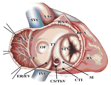
TheBesian Valve
The coronary sinus contains different valves, and the most popular valve is the Thebesian valve at the ostium of the coronary sinus. The Thesbesian valve is a calved shaped structure often found shielding the mouth of the coronary sinus as it opens to the right atrium. These valves are extremely erratic and sometimes may present an impediment during cannulation of the coronary sinus.
Coronary sinus also contains some valve like the Valve of Vieussen that always marks the end of the vein. These venous valves are also regularly found at the entrance of the ventricular veins into the great cardiac vein. This can also be seen in the sinus tarsi anatomy, dural venous sinus anatomy, dural sinus anatomy, and also nasal sinus anatomy
The length of the Coronary Sinus
The length of the Coronary Sinus differs from 3 to 5.5 centimeter, and this length depends on the site of the drainage of the posterolatteral vein. The diameter also varies and it dependent on the loading presence, conditions and extent of atrial myocardium with coronary vein, and the presence of underlying cardiac disease or prior cardiac surgery.
Positioning of the Coronary Sinus
The Coronary Sinus lies in the sulcus between the left atrium and ventricle and is a continuation of the great cardiac vein from the valve of the great cardiac vein to the ostium of the Coronary Sinus as it terminates in the right atrium. The Coronary Sinus initiated at the right atrial orifice and also ends at the valve of Vieussen’s. The coronary sinus collects blood from the ventricular veins during ventricular systole and unfilled into the atrium during atrial systole.
The wall of the coronary system is made up of striated myocardium that is incessant with atria, forming a myocardial sleeve around the venous system. Normally, this sleeve does not broaden into the ventricle myocardium. In contrast, where a continuation into the ventricle is formed, however, an atrioventricular bypass tract referred to as an epicardial pathway is formed.
Conclusively, having an accurate knowledge of the, sinuses anatomy is extremely important. In this article today, we explain all about coronary sinus, cavernous sinus anatomy, the development, length and positioning of the coronary sinus, so as to improve your understanding.
Sources and Citations: stop-sinusitis.net
Leave a Reply